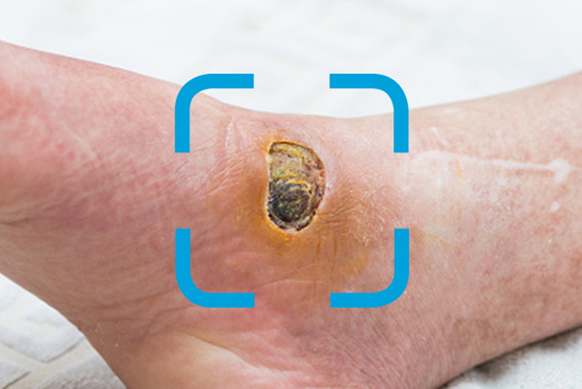
Arterial vs venous ulcers: What’s the difference?
Use this guide to properly identify two common lower extremity wounds.

Lower extremity wounds can be tricky to identify, as they have many similarities. However, some key characteristics will help you correctly identify them. The most common types of lower extremity wounds are venous ulcers and arterial ulcers.
In this article, we’ll explore the differences between venous leg ulcers and arterial, or ischemic, ulcers. Knowing their key features, such as location and size, can help you determine proper wound care and improve patient outcomes.
.
Characteristics of venous leg ulcers
Location: Venous leg ulcers usually develop on the inner lower leg, above the medial malleolus, gaiter area.
Size and shape: Wounds are often shallow, but large, and typically have irregular edges that may also slope.
Color: Typically, venous wounds appear ruddy red, with granular tissue. There may also be discoloration with yellow slough present.
Appearance: Surrounding skin may be shiny, warm or scaly. Tunneling is uncommon.
Exudate: These wounds often appear “wet,” as they often have moderate to heavy exudate, requiring absorbent wound care dressings.
Pain level: Individuals often describe a dull, aching pain. This pain is more likely due to the underlying chronic venous insufficiency and resulting edema rather than from the wound itself.
Other facts and tips about venous ulcers:
- In the case of infection, there is often an accompanying foul odor, and may be purulent.
- Venous ulcers are the most common form of lower extremity wound, accounting for 80% – 90% of all leg ulcers.
- Venous leg ulcers frequently recur. That’s why it’s important to treat the underlying venous disease with compression and other strategies.

Characteristics of arterial wounds
Location: Arterial wounds, caused by a lack of blood circulation, occur most often on the foot, in between or at the tips of the toes, at pressure points from foot wear, on the heels and around lateral malleolus (the bone on the outside of the ankle joint).
Size and shape: Most likely round, with a “punched out” appearance, they may range in size from small to large, with well-defined edges.
Color: Often occur yellow, brown or black in color. Skin may also appear pale and non-granulating.
Appearance: Arterial ulcers are often deep, but may also appear shallow in early stages. Skin surrounding the wound is often thin, smooth, taut and dry. Loss of hair on the leg is also common.
Exudate: Unlike venous ulcers, arterial ulcers are often dry due to minimal drainage.
Pain level: Reportedly very painful. Elevating the leg can increase this pain.
Other distinguishing characteristics:
- Toenails often appear brittle, yellow, deformed, thick and dry.
- A patient’s pulse may be indistinguishable around the site of the wound.
- The area around the wound is likely cool or cold to the touch due to minimal blood circulation.
Learn more about venous ulcers and preventing skin breakdown:
Download these educational posters to help distinguish lower extremity wounds
Explore new ways to help patients manage venous stasis disease
Make it easier for caregivers to do the right thing with a holistic approach to skin health


